


 2 / 4
2 / 4

3-D
3-D
{
What is 3-D mammography?
}
It’s an x-ray technology that takes
multiple images of the breast from dif-
ferent angles. The images are combined
to create a three-dimensional picture so
radiologists can examine breast tissue
one thin layer at a time.
“Compared to traditional mammog-
raphy, it’s like the difference between
looking only at the cover of a book and
opening the book to read each page,”
Hull says.
{
How are 3-D pictures better?
}
“The goal of mammography is
to find breast cancer when it’s as
small as possible,” Hull says. “And 3-D
mammography is exceptional for find-
ing very small abnormalities.” It also
makes detecting cancer in dense breast
tissue easier.
{
Is the actual screening any
different?
}
“A 3-D mammogram takes a few
seconds longer than 2-D—which women
probably won’t notice,” Hull says. Other-
wise, patient prep is the same, the equip-
ment looks the same and the breasts are
still compressed for imaging.
{
Is 3-D the only option now?
}
No. Your doctor may feel that 2-D
mammography is fine for you. “If you’re
older and don’t have dense breasts, maybe
you don’t need 3-D,” Hull says. She notes
that, in general, the younger a woman is,
the denser her breasts tend to be.
{
What about cost?
}
3-D screening costs $50 more than
a regular mammogram. Patients pay
only that amount out of pocket. If their
insurance covers 3-D imaging, GVMH
reimburses them later.
“Patients today are much more edu-
cated about their health and what’s avail-
able,” Hull says. “And they were asking,
‘When are you getting 3-D mammogra-
phy?’ We’re excited to offer this important
new service.”
Women who get timely mammograms are en-
listing a powerful tool to help guard against
breast cancer.
These tests can help spot breast cancer
early—long before it causes symptoms—
when treatment often works best.
Starting at age 40, have a yearly mammo-
gram along with breast exams done by your
doctor, urges the American Cancer Society
(ACS).
After your mammogram, a radiologist
will examine your images for any abnormal
areas.
These images can reveal tissue changes,
including:
Calcifications.
These mineral deposits
appear as white spots on a mammogram.
Larger spots are usually harmless
changes, often related to aging.
However, groups of tiny, white specks are
sometimes signs of cancer.
Masses.
Various lumps and masses may
also be found, including fluid-filled cysts or
other noncancerous growths. The size, shape
and edges of a mass can be important.
For example, noncancerous masses often
have well-defined edges, rather than irregular
ones.
Think positive
If something suspicious is found, try not
to worry too much. In the vast majority of
cases, it isn’t cancer.
More testing—such as another mam-
mogram, an ultrasound or a biopsy—may be
needed.
According to the ACS, less than 10 per-
cent of women who are called back for more
tests are found to have breast cancer.
The bottom line: Screening for breast
cancer can provide peace of mind and help
protect your health.
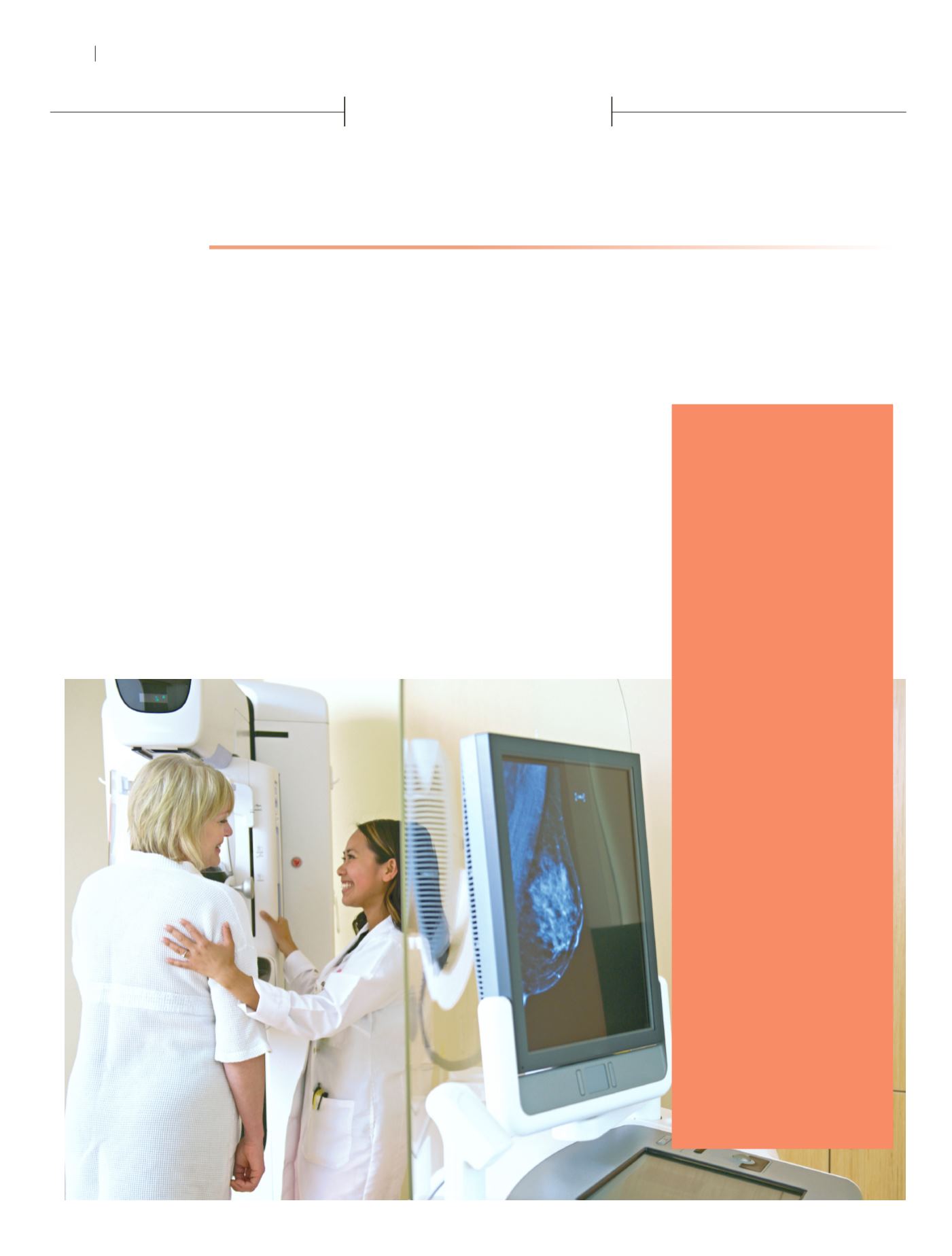
What doctors look
for when reading a
mammogram
MAMMOGRAMS NOW AT GVMH
Ready for your
mammogram? Call
660-890-7145 to
make an appointment.
Mammography is the best tool doctors have to screen for breast cancer. Now Golden Valley Memorial
Hospital (GVMH) offers the best of the best: 3-D mammography.
✦
“Breast tomosynthesis—or
3-D mammography—is the most advanced technology available for breast imaging,” says Tami
Hull, Director of Imaging Services at GVMH. “So it was an excellent tool to add to our breast cancer
screening.” Here, Hull helps answer questions about this new service.
B r e a s t C a n c e r S c r e e n i n g
2
W I N T E R 2 0 1 6
H E A L T H
S C E N E