


 2 / 4
2 / 4

We l l n e s s
—Continued from front page
H
IGH-TECH,
H
IGH-QUALITY CARE
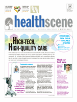
3-D digital mammography
(breast tomosynthesis)
Getting a 3-D mammogram is a lot like getting a regular 2-D mammogram. But with the 3-D
version, the x-ray tube moves across the breast, taking images from different angles that are
reassembled as three-dimensional slices—similar to a CT scan.
“Think of a book,” explains Tami Hull, Director of Imaging Services at GVMH. “You see
the front cover. But as you flip through it, you see all the individual pages separately. In a
similar way, we get multiple images on a 3-D mammogram that allow us to see different
layers of breast tissue.”
■
■
How it helps:
3-D mammography may help radiologists better detect breast cancer.
And because of the multiple views of breast tissue, it may also reduce the number of false
positives—suspicious findings that trigger additional tests, which ultimately determine
everything is OK.
■
■
Available:
Monday through
Friday, 7 a.m. to 5:30 p.m.
■
■
Purchased:
Fall 2015
Nuclear medicine heart scan
The new D-SPECT equipment at GVMH provides a reveal-
ing look at how the heart functions. For this test, a safe
radioactive tracer is injected into the bloodstream. The
tracer travels to the heart and releases energy. Cameras
detect the released energy and use it to create pictures of
the heart.
■
■
How it helps:
This test can reveal things like damaged
heart muscle from a previous heart attack or blood-flow
problems that may cause heart angina (chest pain). The
results can help doctors determine treatment.
“Our new equipment produces better pictures, takes
less time and is much more accurate than our old equip-
ment,” says Tami Hull, Director of Imaging Services at
GVMH. “It’s known as the best in the market at producing
these images.”
■
■
Available:
Monday through Friday, 7 a.m. to 4:30 p.m.
■
■
Purchased:
Spring 2016
“We get multiple images
on a 3-D mammogram
that allow us to see
different layers of
breast tissue.”
—Tami Hull,
Director of Imaging Services at GVMH
“Our new
equipment
produces better
pictures, takes
less time and
is much more
accurate than
our old equipment.”
—Tami Hull,
Director of Imaging Services at GVMH
WHEN YOU NEED
an imaging test, your doctor has a number of technologies to turn to. And,
although it’s been around the longest, doctors still rely on the x-ray.
Medical x-rays are a fast, potentially lifesaving tool for detecting injuries and illnesses. They can
reveal broken bones, tumors, heart disease, kidney stones, pneumonia and many other conditions.
Other imaging methods have evolved from the x-ray. CT scans and mammography, for example,
both use x-ray technology.
SAFETY CONCERNS
To create images, x-rays produce radiation. While special care is taken to
use the lowest amount possible, the body does absorb some radiation.
However, the benefits of an accurate diagnosis generally outweigh any risk from radiation,
according to the U.S. Food and Drug Administration.
Women should inform their doctor if they are or could be pregnant. If an x-ray is still necessary,
an x-ray technologist can use special precautions or take fewer x-rays than are normally required.
Always feel free to discuss your concerns about radiation from x-rays, or any other source,
with your doctor.
Additional source: Radiological Society of North America
X-RAYS
STILL EFFECTIVE—AND STILL SAFE
2
W I N T E R 2 0 1 7
H E A L T H
S C E N E